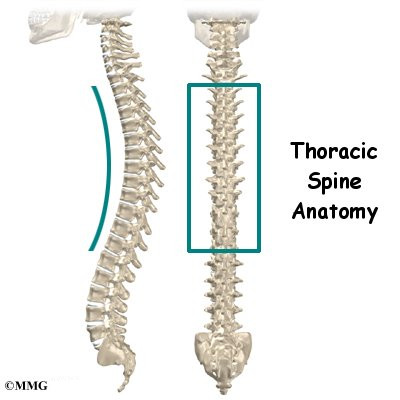
A thoracic spine MRI scan examines the middle section of your spine between the neck and lower back.

Magnetic resonance imaging of the thoracic spine: Part 1: normal imaging anatomy - ScienceDirect

Blind Spots in Spine Imaging • APPLIED RADIOLOGY

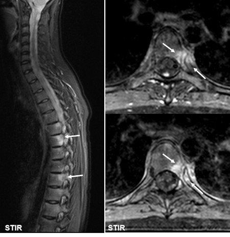
Frontiers Successful treatment of thoracic myelopathy caused by spontaneous spinal epidural hematoma (SSEH) combined with calcification of the ligamentum flavum (CLF) by posterior percutaneous endoscopic surgery (PPES): A case report

How to Read a MRI of the Normal Thoracic Spine (Mid Back)

Normal thoracic spine MRI, Radiology Case
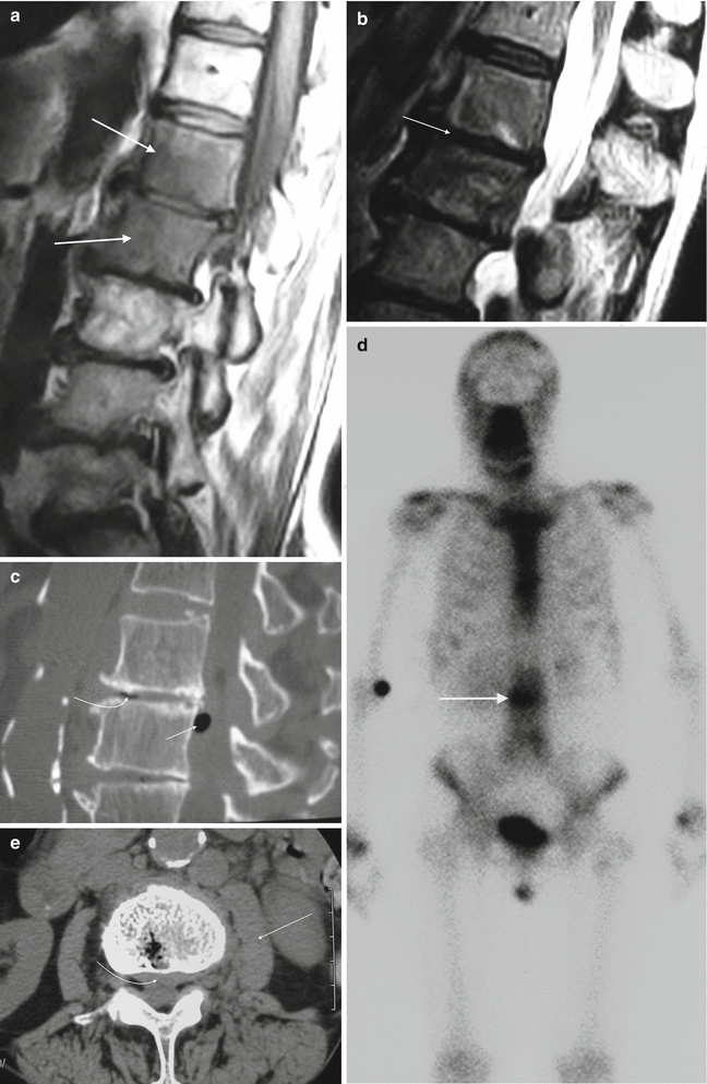
Frontiers Multiple ossified spinal meningiomas in the thoracic spine: A case report and literature review

Thoracic Spine MRI. 37year old female. Got MRI back but was told inconclusive. Have been having right upper back pain and excruciating pain when any pressure is applied to the area. Would

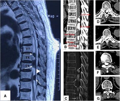
MRI OF THORACIC SPINE HISTORY A 53-year-old woman, presented with paresis and paresthsia of both legs. Longitudinally extensive T2W hyperintensity at spinal cord . 15763120 Stock Photo at Vecteezy

Myelopathy, Dr. Michael Steinhaus, Minimally Invasive Spine Surgeon
Spine Infection Mimics