![](https://www.researchgate.net/publication/322368228/figure/fig4/AS:962647902130176@1606524553424/Three-dimensional-model-of-the-elements-of-the-pelvic-girdle-ie-innominates-femur.gif)
![](https://www.frontiersin.org/files/Articles/1036882/fbioe-10-1036882-HTML/image_m/fbioe-10-1036882-g007.jpg)
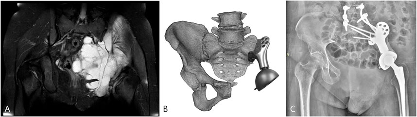
Frontiers Reconstruction with 3D-printed prostheses after type I + II + III internal hemipelvectomy: Finite element analysis and preliminary outcomes
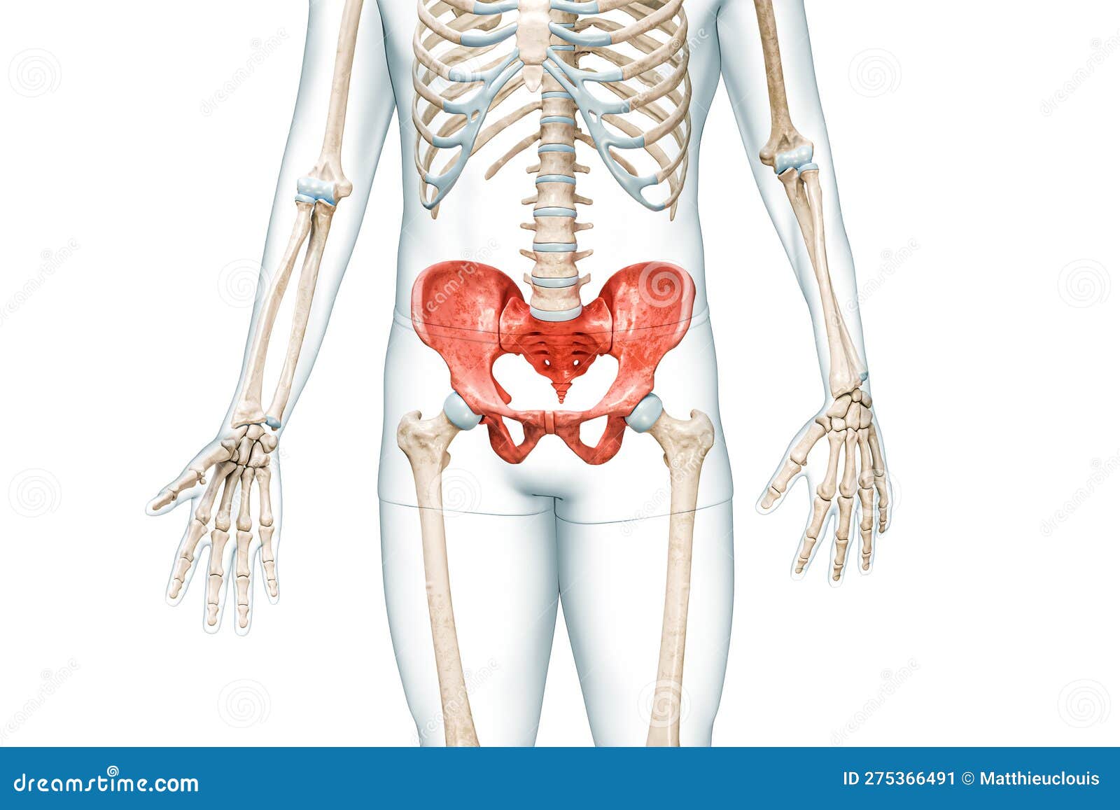
![](https://upload.wikimedia.org/wikipedia/commons/thumb/9/98/Gray241.png/640px-Gray241.png)
Pelvis - Wikipedia
![](https://www.frontiersin.org/files/Articles/541321/fbioe-08-00963-HTML/image_m/fbioe-08-00963-g001.jpg)
Frontiers Determination of Three-Dimensional Corrective Force in Adolescent Idiopathic Scoliosis and Biomechanical Finite Element Analysis
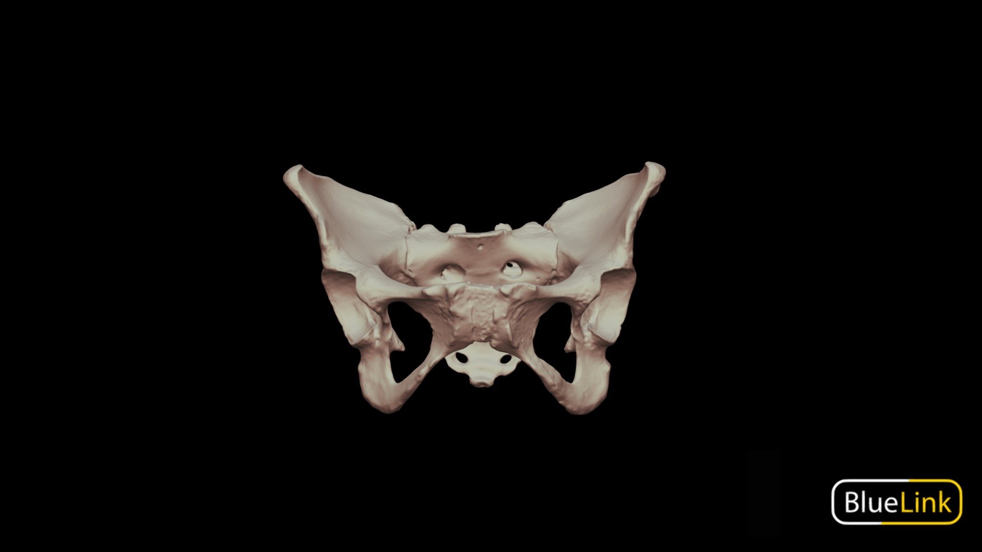
![](https://media.sketchfab.com/models/53240ae515b644d989f0330a9280e5ac/thumbnails/9998ddc49468483b87a41880259433ec/1d532518eba745f5901d4f0755bba140.jpeg)
3D model pelvis
![](https://www.researchgate.net/publication/236690119/figure/fig1/AS:214059065581589@1428047066907/The-three-dimensional-finite-element-model-of-the-hip-joint-A-The-entire-model.png)
The three dimensional finite element model of the hip joint. A–The
![](https://i.ytimg.com/vi/I87G1dUcZng/hq720.jpg?sqp=-oaymwEhCK4FEIIDSFryq4qpAxMIARUAAAAAGAElAADIQj0AgKJD&rs=AOn4CLDf_FpKNEa3skr3e6RfhclI81EqjQ)
Bones of the Pelvis (3D Anatomy Tutorial), UKMLA
![](https://c5.rgstatic.net/m/435982309481010/images/template/default/author/author_default_m.jpg)
PDF) Shape Covariation (or the Lack Thereof) Between Vertebrae and
![](https://teachmeanatomy.info/wp-content/uploads/Overview-of-the-Pelvic-Cavity-and-the-Pelvic-Floor-Muscles.jpg)
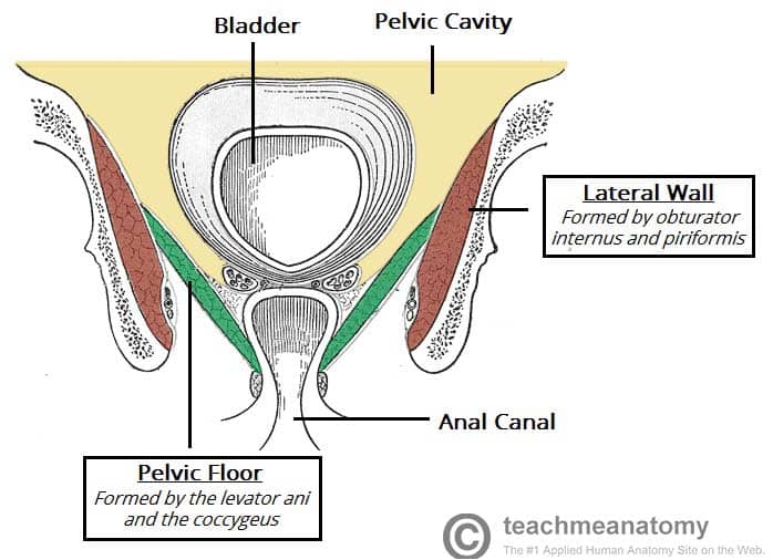
The Pelvic Floor - Structure - Function - Muscles - TeachMeAnatomy
![](https://www.researchgate.net/publication/322368228/figure/fig3/AS:962647897956355@1606524552918/Three-dimensional-model-of-the-elements-of-the-pectoral-girdle-scapula-and-forelimb_Q320.jpg)
Three-dimensional model of the elements of the pelvic girdle (i.e.
![](https://www.frontiersin.org/files/Articles/698179/fsurg-08-698179-HTML/image_m/fsurg-08-698179-g001.jpg)
Frontiers A Novel Three-Dimensional Computational Method to Assess Rod Contour Deformation and to Map Bony Fusion in a Lumbopelvic Reconstruction After En-Bloc Sacrectomy
![](https://www.frontiersin.org/files/Articles/527766/fbioe-08-00318-HTML/image_m/fbioe-08-00318-g001.jpg)
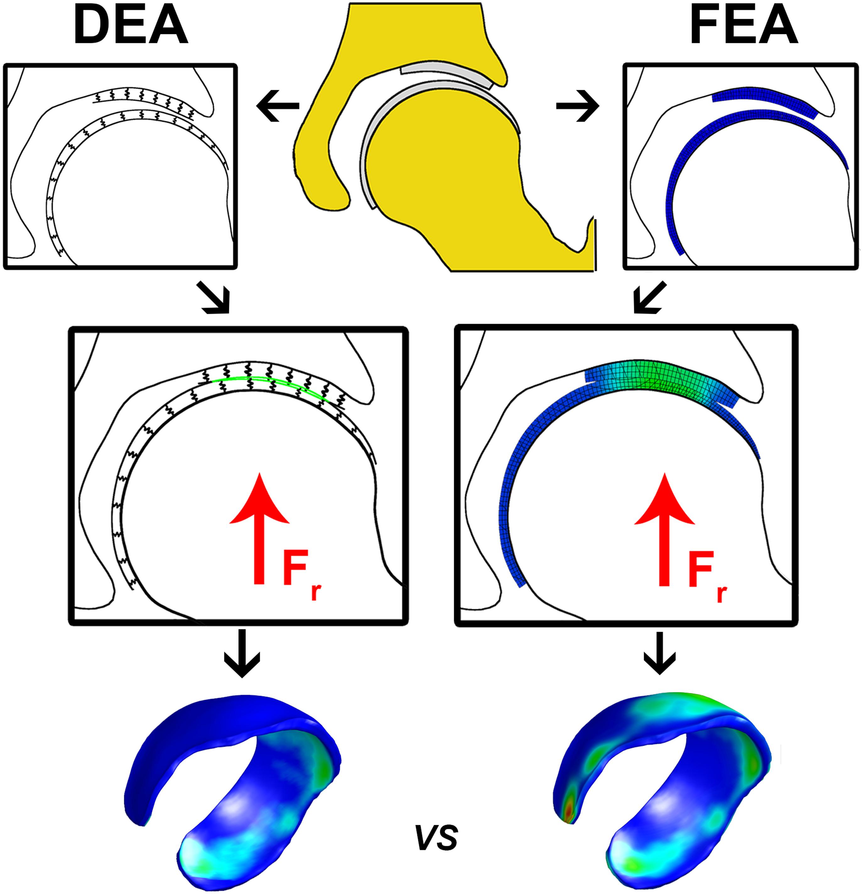
Frontiers A Combined Geometric Morphometric and Discrete Element Modeling Approach for Hip Cartilage Contact Mechanics
![](https://www.kenhub.com/thumbor/ERJ2NXsLu07ICkotydo39yd9Qxg=/fit-in/800x1600/filters:watermark(/images/logo_url.png,-10,-10,0):background_color(FFFFFF):format(jpeg)/images/library/13792/Bony_pelvis__Pelvis_ossea_.png)
Bony pelvis: Ilium, ischium, pubis
![](https://c8.alamy.com/comp/RN7PX6/the-anatomy-of-the-domestic-animals-veterinary-anatomy-the-pelvis-111-diameter-transversa-is-measured-at-the-greatest-width-i-e-just-above-the-psoas-tubercle-the-posterior-aperture-igtr-outlet-of-the-pelvis-apertura-pelvis-caudalis-is-mueh-smaller-and-is-very-incomplete-in-the-skeleton-it-is-bounded-dorsaily-by-the-fig-s9pelvic-bones-of-staluon-front-view-c-conjugate-dt-transverse-diameter-of-pelvic-inlet-please-note-that-these-images-are-extracted-from-scanned-page-images-that-may-have-been-digitally-enhanced-for-readability-coloration-and-appearance-of-these-i-RN7PX6.jpg)
The anatomy of the domestic animals. Veterinary anatomy. THE PELVIS 111 (Diameter transversa) is measured at the greatest width, i. e., just above the psoas tubercle. The posterior aperture i>r outlet
![](https://upload.wikimedia.org/wikipedia/commons/thumb/f/f9/1116_Muscle_of_the_Female_Perineum.png/350px-1116_Muscle_of_the_Female_Perineum.png)
Pelvis - Wikipedia
![](https://media.springernature.com/m685/springer-static/image/art%3A10.1007%2Fs11692-017-9443-6/MediaObjects/11692_2017_9443_Fig7_HTML.gif)
Shape Covariation (or the Lack Thereof) Between Vertebrae and